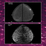
Introduction to Contrast-Enhanced Mammography
Contrast-Enhanced Mammography (CEM) is an advanced imaging technique used in radiology to enhance the visualization and detection of breast abnormalities, particularly in women with dense breast tissue or those at high risk for breast cancer. This innovative method combines the benefits of traditional mammography with the administration of a contrast agent, resulting in improved diagnostic accuracy and earlier detection of malignancies.
How CEM Works
CEM involves the injection of an iodine-based contrast agent into the patient’s bloodstream prior to performing a mammogram. The contrast agent highlights areas of increased blood flow, which is often associated with malignant tumors due to their increased vascularity. By comparing the contrast-enhanced images with standard mammography images, radiologists can better identify and characterize suspicious lesions.
The procedure typically involves the following steps:
- A standard mammogram is performed to obtain baseline images of the breasts.
- An intravenous line is inserted into the patient’s arm, and the contrast agent is administered.
- After a short waiting period to allow the contrast to circulate, additional mammographic images are acquired.
- The contrast-enhanced images are then compared with the baseline images to identify any areas of abnormal enhancement.
Advantages of CEM
CEM offers several advantages over traditional mammography and other breast imaging modalities:
-
Improved sensitivity: CEM has been shown to have higher sensitivity in detecting breast cancers, particularly in women with dense breast tissue, where traditional mammography may have limited effectiveness.
-
Reduced false positives: By providing additional information about the vascularity of lesions, CEM can help reduce the number of false-positive findings, leading to fewer unnecessary biopsies and patient anxiety.
-
Cost-effective: CEM is a relatively low-cost alternative to more expensive imaging modalities such as breast MRI, making it more accessible to a wider range of patients.
-
Faster acquisition: CEM exams can be performed in a shorter time compared to breast MRI, reducing patient discomfort and increasing throughput in the radiology department.
Indications for CEM
CEM is particularly useful in the following clinical scenarios:
-
Dense breast tissue: Women with dense breast tissue have a higher risk of developing breast cancer, and traditional mammography may have limited sensitivity in detecting abnormalities. CEM can provide additional information to help identify cancers that might otherwise be missed.
-
High-risk patients: Women with a family history of breast cancer, genetic mutations (e.g., BRCA1 or BRCA2), or other risk factors may benefit from the enhanced detection capabilities of CEM.
-
Inconclusive findings: When traditional mammography or ultrasound results are inconclusive or equivocal, CEM can provide additional information to help characterize lesions and guide management decisions.
-
Monitoring response to treatment: CEM can be used to evaluate the response of breast cancers to neoadjuvant chemotherapy, helping to assess the effectiveness of treatment and guide further management.
Limitations and Risks
While CEM offers many benefits, it is important to consider its limitations and potential risks:
-
Radiation exposure: CEM involves additional radiation exposure compared to standard mammography due to the acquisition of multiple images. However, the overall radiation dose is still relatively low and considered safe for most patients.
-
Contrast agent reactions: As with any procedure involving contrast agents, there is a small risk of allergic reactions or adverse effects. Patients with known allergies to iodine or contrast agents should inform their healthcare provider before undergoing CEM.
-
False negatives: Although CEM has improved sensitivity compared to traditional mammography, it is not perfect and may still miss some cancers, particularly those that do not exhibit increased vascularity.
-
Limited availability: CEM is a relatively new technology and may not be widely available in all healthcare facilities, particularly in smaller or rural settings.

Comparison with Other Imaging Modalities
CEM is one of several imaging modalities used for breast cancer detection and characterization. It is important to understand how CEM compares to other techniques:
CEM vs. Traditional Mammography
Traditional mammography remains the gold standard for breast cancer screening and is widely available and cost-effective. However, it has limitations in detecting cancers in dense breast tissue and may result in a higher number of false positives. CEM builds upon the foundation of traditional mammography by adding the contrast enhancement component, leading to improved sensitivity and specificity in certain patient populations.
CEM vs. Breast Ultrasound
Breast ultrasound is often used as a complementary tool to mammography, particularly in women with dense breast tissue or for the evaluation of palpable lumps. Ultrasound is non-invasive, widely available, and does not involve radiation exposure. However, it is operator-dependent and may not be as effective in detecting small or non-palpable cancers. CEM can provide additional information that ultrasound may not be able to capture, particularly regarding the vascularity of lesions.
CEM vs. Breast MRI
Breast MRI is considered the most sensitive imaging modality for detecting breast cancers, particularly in high-risk patients or those with dense breast tissue. It provides excellent soft tissue contrast and can help characterize lesions based on their enhancement patterns. However, breast MRI is more expensive, time-consuming, and may have a higher false-positive rate compared to other modalities. CEM offers a more cost-effective and faster alternative to breast MRI, with comparable sensitivity in some patient populations.
| Imaging Modality | Advantages | Limitations |
|---|---|---|
| Traditional Mammography | – Widely available – Cost-effective – Gold standard for screening |
– Limited sensitivity in dense breasts – Higher false-positive rate |
| Breast Ultrasound | – Non-invasive – No radiation exposure – Useful for palpable lumps |
– Operator-dependent – May miss small or non-palpable cancers |
| Breast MRI | – Highest sensitivity – Excellent soft tissue contrast – Useful for high-risk patients |
– Expensive – Time-consuming – Higher false-positive rate |
| CEM | – Improved sensitivity in dense breasts – Reduced false positives – Cost-effective alternative to MRI |
– Additional radiation exposure – Contrast agent reactions – Limited availability |
Clinical Applications and Case Studies
CEM has been increasingly used in various clinical scenarios to improve breast cancer detection and characterization. Several studies have demonstrated its effectiveness in specific patient populations and clinical settings.
Case Study 1: Dense Breast Tissue
A 45-year-old woman with heterogeneously dense breast tissue presented for routine screening mammography. The mammogram showed no apparent abnormalities. However, given her dense breast tissue, a CEM exam was performed. The contrast-enhanced images revealed a small, enhancing lesion in the upper outer quadrant of the right breast, which was not visible on the standard mammogram. Subsequent biopsy confirmed the presence of an invasive ductal carcinoma. The early detection enabled by CEM allowed for prompt treatment and improved patient outcomes.
Case Study 2: High-Risk Patient
A 35-year-old woman with a strong family history of breast cancer and a known BRCA1 mutation presented for screening. Given her high-risk status, a CEM exam was performed in addition to standard mammography. The CEM images identified a suspicious enhancing lesion in the left breast, which was not apparent on the mammogram. Biopsy revealed the presence of a high-grade ductal carcinoma in situ (DCIS). The early detection and characterization of the lesion through CEM allowed for appropriate management and prevention of progression to invasive cancer.
These case studies highlight the potential of CEM to improve breast cancer detection and characterization in specific patient populations, leading to earlier diagnosis and better outcomes.
Future Directions and Research
As CEM continues to gain recognition in the radiology community, ongoing research aims to further evaluate its effectiveness and expand its applications:
-
Large-scale studies: Additional large-scale, multi-center studies are needed to validate the findings of initial research and establish the role of CEM in various clinical scenarios.
-
Cost-effectiveness analysis: While CEM is considered a cost-effective alternative to breast MRI, more comprehensive economic analyses are necessary to assess its long-term impact on healthcare costs and resource utilization.
-
Technological advancements: Improvements in CEM technology, such as enhanced contrast agents or advanced image processing techniques, may further enhance its diagnostic accuracy and clinical utility.
-
Combinatorial approaches: Researchers are exploring the potential of combining CEM with other imaging modalities or biomarkers to develop more personalized and targeted screening and diagnostic strategies.
As research progresses, it is anticipated that CEM will play an increasingly important role in the early detection and management of breast cancer, ultimately improving patient outcomes and quality of life.
Frequently Asked Questions (FAQs)
-
What is Contrast-Enhanced Mammography (CEM)?
Contrast-Enhanced Mammography (CEM) is an advanced imaging technique that combines traditional mammography with the administration of a contrast agent to improve the visualization and detection of breast abnormalities, particularly in women with dense breast tissue or those at high risk for breast cancer. -
How does CEM differ from traditional mammography?
CEM builds upon the foundation of traditional mammography by adding a contrast enhancement component. After a standard mammogram is performed, an iodine-based contrast agent is injected into the patient’s bloodstream, and additional images are acquired. The contrast agent highlights areas of increased blood flow, which is often associated with malignant tumors, leading to improved sensitivity and specificity in detecting breast cancers. -
Who can benefit from CEM?
CEM is particularly useful for women with dense breast tissue, high-risk patients (e.g., those with a family history of breast cancer or genetic mutations), cases with inconclusive findings on traditional mammography or ultrasound, and for monitoring response to breast cancer treatment. -
Are there any risks associated with CEM?
CEM involves additional radiation exposure compared to standard mammography, although the overall dose is still relatively low and considered safe for most patients. There is also a small risk of allergic reactions or adverse effects related to the contrast agent. Patients with known allergies to iodine or contrast agents should inform their healthcare provider before undergoing CEM. -
How does CEM compare to other breast imaging modalities?
CEM offers improved sensitivity and reduced false positives compared to traditional mammography, particularly in women with dense breast tissue. It is a cost-effective and faster alternative to breast MRI, with comparable sensitivity in some patient populations. CEM can provide additional information that breast ultrasound may not capture, particularly regarding the vascularity of lesions.
Conclusion
Contrast-Enhanced Mammography (CEM) is a promising advancement in breast imaging, offering improved sensitivity and specificity in detecting breast cancers, particularly in women with dense breast tissue or those at high risk. By combining the benefits of traditional mammography with the administration of a contrast agent, CEM provides valuable information about the vascularity of lesions, leading to earlier detection and better characterization of abnormalities.
While CEM has its limitations and may not replace other imaging modalities entirely, it serves as a valuable tool in the radiologist’s arsenal for breast cancer screening and diagnosis. As research continues to validate its effectiveness and expand its applications, CEM is poised to play an increasingly important role in improving patient outcomes and quality of life.
Ultimately, the decision to use CEM should be made on a case-by-case basis, considering the individual patient’s risk factors, breast density, and clinical history. By working closely with healthcare providers and staying informed about the latest advances in breast imaging, patients can ensure that they receive the most appropriate and effective care for their unique needs.

No responses yet